|
SCREENING
Unique tool for screening purposes
PRINCIPLE
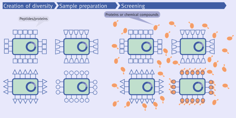
A unique feature of the autodisplay system is its application as a tool for screening purposes. This challenging application is basically comprised of three steps: (i) cell surface display of peptide libraries, (ii) labeling of single cells with an inhibitor structure by target protein binding, and (iii) selecting and sorting of labeled cells by fluorescence activated cell sorting (FACS). The rationale of this concept is based on the idea that a typical inhibitor has a high affinity for its target and vice versa. The affinity of a given target protein to an inhibitor can be exploited by using specific E. coli cells displaying an active inhibitor at the cell surface. If the target protein is coupled to a fluorescent dye, flow cytometry can be used to sort single cells expressing an inhibitor with affinity for the target protein. The selected cells can subsequently be used for clonal production of cell quantities sufficient for analytical or preparative purposes. This concept takes advantage of the high copy number of each molecule expressed on the E. coli cell surface using the autodisplay system and of the good viability and integrity of these cells. The first advantage enhances the selection of positive from negative variants, and the latter is crucial for the number of clones surviving the FACS sorting.
 |
SELECTED EXAMPLES
Cathepsin G: Human cathepsin G is a serine proteinase that accumulates in the human azurophil granule of neutrophils. Under certain circumstances as found in inflammatory diseases the proteinase can participate in local destruction of connective tissue proteins. Therefore, cathepsin G inhibitors are promised to be an important tool in the treatment of chronic inflammatory diseases such as rheumatoid arthritis or pulmonary emphysema. On the basis of the known peptidic cathepsin G inhibitor P15, a random library using autodisplay was constructed, in which every E. coli cell displayed a different variant of P15. To identify new inhibitory lead structures this library was screened against fluorescently labeled cathepsin G, based on the idea that E. coli cells expressing an enzyme inhibitor at the surface can be specifically labeled by its target and identified using fluorescence activated cell sorting (FACS). Using this approach it was possible to identify different new inhibitory lead structures directed against cathepsin G with the best lead structure showing an IC50 of 11.7 µM.
Antibody fragments: Autodisplay can also be used for the display of antibody fragments on the surface of E. coli. One possibility is the expression of the light chain (VL) and the heavy chain (VH) as one fusion protein connected with a short linker [single chain variable fragments (scFv)]. The mobility of the autodisplay anchoring domain in the outer membrane also enables the expression of the VH and VL independently of each other. Both genes encoding the VH and VL are localized on different plasmids in the same cell. They are expressed in a monomeric state and aggregate spontaneously on the surface of E. coli to a functional antibody fragment (twin chain variable fragment (tcFv): E. coli cells displaying scFv or tcFv fragments directed against epidermal growth factor receptor were able to bind specifically on tumor cells displaying epidermal growth factor receptor.
|